
TPLO planning radiographs
Coordinate new planning radiographs with the surgery team. Use the positioning and calibration examples below when taking the views at your clinic.
Positioning examples
Print TPLO positioning + example radiographs · sheet 1
Dr. Wood plans each TPLO in VPOP from calibrated radiographs. Positioning or calibration errors may require repeat views.
Calibration ball on EVERY radiograph.
Sedation or anaesthesia is often needed. Include a calibration marker and a left/right marker in each view.
True lateral view
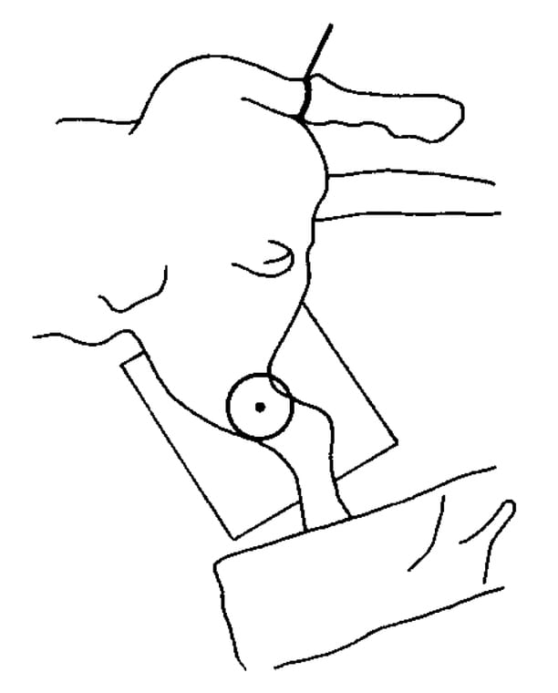
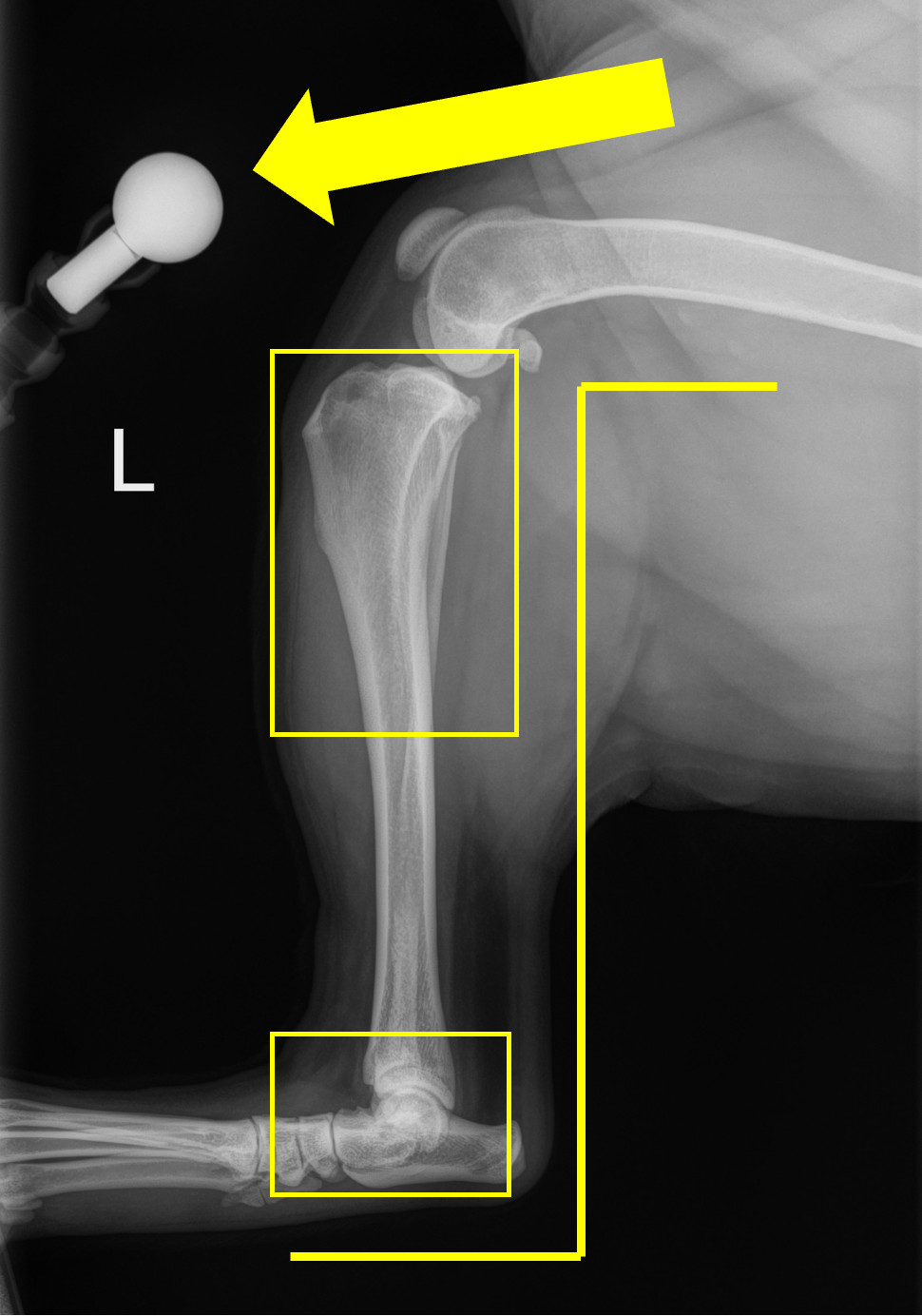
- Stifle and hock near 90 degrees.
- Hock included; beam centred on the stifle/proximal tibia.
- Femoral condyles/fabellae superimposed as well as possible.
Orthogonal CrCd/AP view
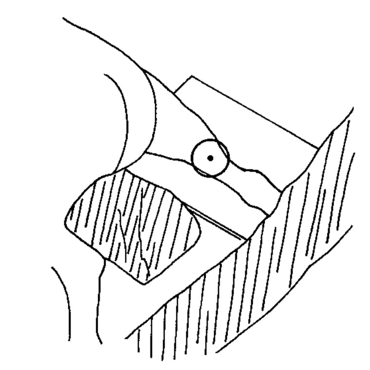
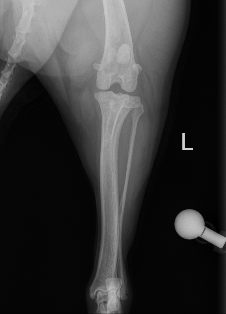
- Limb straight with patella centred.
- Fabellae bisected and hock/calcaneus aligned.
- Stifle, tibia, and hock included.
Coverage and positioning comparisons
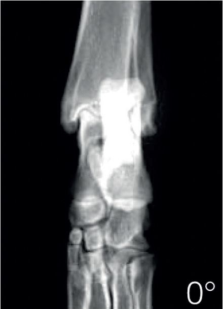
Use the 0° inset to check neutral alignment of the hock (tarsus) on the CrCd view.
 Open hock image ↗
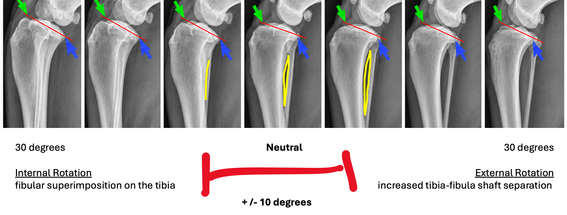
Internal rotation increases overlap of the fibula and tibia. External rotation increases their separation.
 Open rotation strip ↗
The red bracket marks ±10° around neutral in the teaching strip. In a cadaver study of CrCL-deficient stifles without osteoarthritis, up to 30° of tibial rotation in either direction had no clinically relevant effect on lateral tibial plateau angle (TPA) measurements. Aim for a true lateral. Check coverage and calibration separately. Buirkle et al., 2019.
Examination notes
- Send drawer and tibial thrust findings under sedation or anaesthesia, if available.
- Negative or equivocal awake drawer/thrust does not rule out CCL disease; a painful or guarded dog may need re-examination.
Discuss with the owner
- TPLO may be an option for a painful, unstable knee caused by cranial cruciate ligament (CCL) disease. It changes how the knee bears weight rather than replacing the ligament.
- The surgical plan is confirmed after specialist examination and imaging review.
- A meniscal click supports suspected injury; no click does not rule out a tear.
- Coordinate calibrated planning radiographs and current pre-op bloodwork before surgery. Confirm the expected hospital stay with the surgical team.
- For both knees, discuss combined versus staged surgery, hospital stay and home support.