
Articular cartilage zones, matrix and defect response
Read the draft explanation
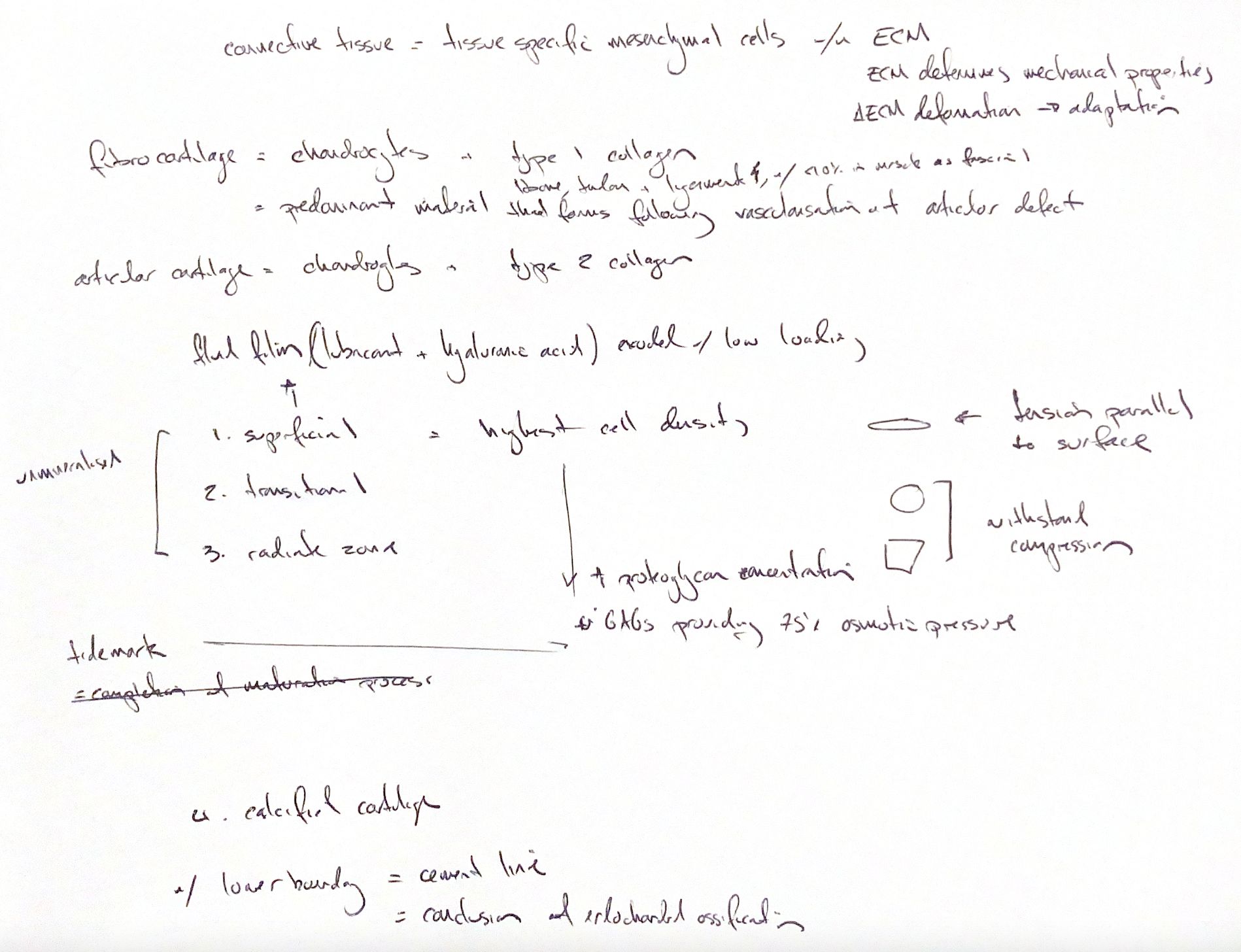
Connective tissue has tissue-specific mesenchymal cells in an extracellular matrix, or ECM. Its matrix gives the tissue its mechanical properties. Loading deforms that matrix and prompts cells to adapt. Skeletal muscle has much less ECM, generally under 10%. Its type I-rich myofascia still carries force.
Articular hyaline cartilage contains chondrocytes and mainly type II collagen. The adult superficial zone has the most cells. Its flat cells and fibrils lie along the surface and resist tension. Transitional cells are rounder. Radiate cells are larger and align across the depth. These three zones are unmineralised. Deeper matrix contains more proteoglycan and helps resist compression.
The tidemark separates unmineralised from calcified cartilage. The cement line sits below mature calcified cartilage. It forms when endochondral ossification of the articular-epiphyseal growth plate ends at maturity.
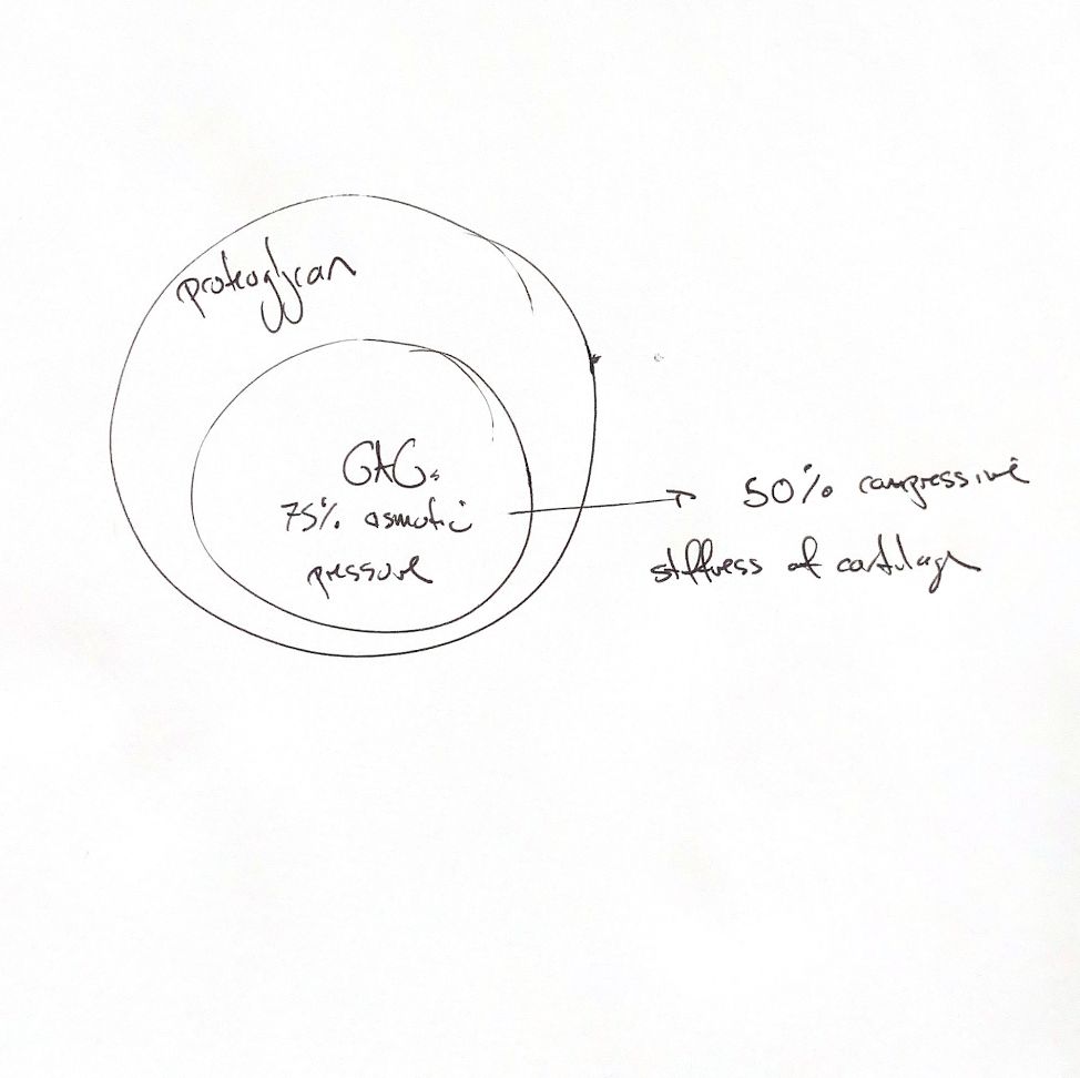
Glycosaminoglycans, or GAGs, draw ions and water into the matrix. Collagen tension restrains that swelling. Classic estimates put GAGs at about 75% of proteoglycan osmotic pressure. Osmotic pressure may supply up to 50% of cartilage compressive stiffness. These are matrix estimates from human-cartilage work and theory, not fixed canine values.
At low loading rates, fluid released from cartilage can form a lubricating film. Lubricin provides boundary lubrication, especially with high load and little motion. Hyaluronic acid provides viscous lubrication. They work together in different modes.
After vascularisation of a full-thickness articular defect, the predominant repair is fibrocartilage. This has chondrocytic cells in a type I-rich matrix. It also forms during healing or degeneration of bone, tendon and ligament.
References
Chris Wood’s study drawings; Johnston & Tobias, Veterinary Surgery: Small Animal, 2nd ed., ch.38, Fig.38.9. Matrix estimates follow its summary of Maroudas et al. 1986 and Lai et al., J Biomech Eng 1991;113:245–258.