
TNM staging and pulmonary imaging
Read the draft explanation
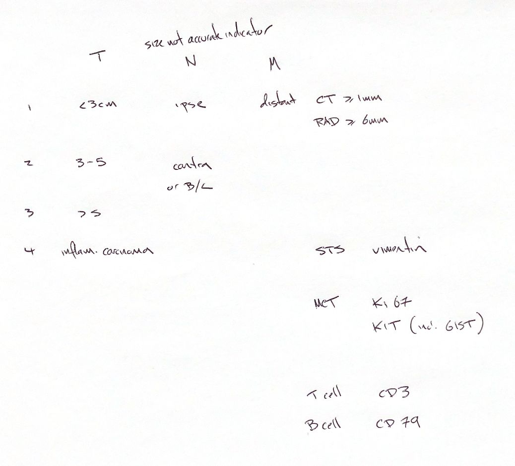
The 3 cm/5 cm size bins and T4 inflammatory carcinoma belong to the modified canine mammary TNM scheme. They should not be used as a general skin-tumour table. T describes the primary tumour, N the regional nodes and M distant metastasis.
For canine mammary tumours, T1 is <3 cm, T2 is 3–5 cm and T3 is >5 cm in maximum diameter. T4 is inflammatory carcinoma. N1 means ipsilateral nodal metastasis; N2 means contralateral or bilateral nodal metastasis. Node size alone cannot establish metastatic status. M0 means no evidence of distant metastasis; M1 means it is detected.
The epidermal/dermal scheme excludes lymphoma, mast cell and mammary tumours. T1 is <2 cm and superficial or exophytic. T2 is 2–5 cm or minimally invasive at any size. T3 is >5 cm or invades subcutis at any size. T4 invades fascia, muscle, bone or cartilage. The node categories also distinguish movable affected nodes from fixed nodes.
Thoracic imaging has a different job: finding pulmonary nodules. In 18 dogs with histologically confirmed pulmonary metastases, 29 paired examinations found an approximate lower detection scale of 1 mm on CT and 7–9 mm for reliable radiographic detection. Some high-detail radiographs can show nodules around 6 mm. Visibility changes with nodule shape, superimposition, lung inflation and technique. Not every CT nodule in that study was individually confirmed as metastatic.
References
Johnston & Tobias, Veterinary Surgery: Small Animal, 2nd ed., Tables 82.2/82.5 and ch. 25; Owen, WHO TNM Classification of Tumours in Domestic Animals, 1980 (modified scheme); Nemanic et al. 2006, JVIM 20:508–515, PMID 16734082.